
Kidney Stones: Water, Climate and When You Need CT KUB
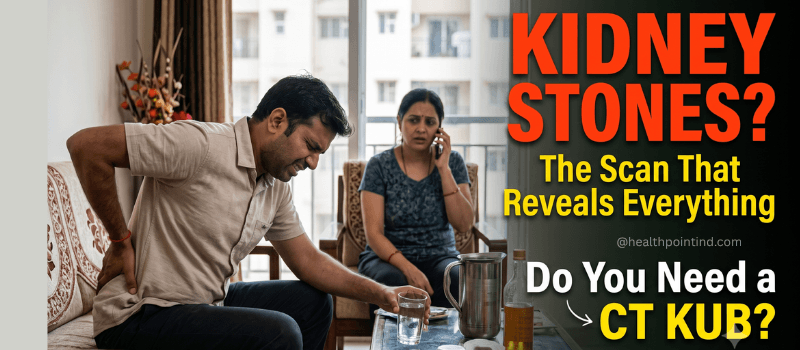
Kidney Stones: Why Water Intake and Climate Matter, and When You Actually Need a CT KUB
Anyone who has experienced renal colic will tell you it is unlike ordinary pain. It arrives suddenly, sits deep in the flank or side of the back, radiates down toward the groin, and comes in waves that no position relieves. People describe pacing, sweating, and being unable to sit still, which is itself diagnostically useful, since most abdominal conditions make people want to lie motionless while a kidney stone makes them restless. What most people do not know when this happens is which scan actually finds the stone, and why the first test they get may not be the one that answers the question.
Why Kidney Stones Form in the First Place
A kidney stone forms when substances in urine become concentrated enough to crystallise and gradually build into a solid mass. The most common type by a considerable margin is calcium oxalate, followed by calcium phosphate, uric acid, struvite, and rarely cystine stones. Each type has slightly different causes and, importantly for imaging, slightly different visibility on different scans.
The single biggest modifiable factor is fluid intake. Dilute urine makes crystallisation far less likely simply because the stone-forming substances never reach the concentration required to precipitate. This sounds almost too simple to be the main driver of a painful condition, but it genuinely is, and it is the reason the first advice any urologist gives after a stone is to substantially increase daily water intake.
The Water Quality and Climate Connection
Two environmental factors influence stone risk in ways that are worth understanding rather than dismissing.
The first is climate and fluid loss. In hot, dry conditions, the body loses considerably more water through sweat and respiration, and urine concentrates as a result. This is why kidney stone incidence tends to rise during summer months and why people working outdoors, or in hot environments, carry higher risk. It is also why many people who feel they drink a reasonable amount of water are still producing concentrated urine, because their losses are higher than they realise.
The second is water hardness. Hard water contains higher levels of dissolved calcium and magnesium. The relationship between hard water and stone formation is more nuanced than commonly assumed, since dietary calcium actually binds oxalate in the gut and can reduce oxalate absorption, and magnesium may have a mildly protective effect. The evidence linking hard water directly to increased stone formation is genuinely mixed rather than settled. What is clearer is that total fluid volume matters more than water composition. Drinking more of whatever water you have access to is more protective than switching water sources while drinking the same amount.
Beyond water, diet contributes meaningfully. High salt intake increases calcium excretion in urine. High animal protein intake raises uric acid and lowers urinary citrate, which normally inhibits stone formation. Oxalate-rich foods including spinach, beetroot, nuts, and tea contribute in people already prone to calcium oxalate stones, though for most people restricting these is less important than increasing fluid.
Symptoms Worth Recognising
Severe, colicky pain in the flank radiating toward the groin is the classic presentation. Blood in the urine, sometimes visible and sometimes only detected on testing, is common. Nausea and vomiting frequently accompany the pain, which occasionally leads to it being mistaken for a gastric problem. Pain or burning during urination, increased urinary frequency, and passing small amounts of urine can occur when a stone sits low in the ureter near the bladder.
Certain features shift this from painful to genuinely urgent. Fever alongside the pain suggests an infected obstructed system, which is a medical emergency requiring immediate attention rather than a wait-and-watch approach. Being unable to pass urine at all, or persistent vomiting preventing any fluid intake, similarly requires prompt medical care.
Which Scan Actually Detects a Kidney Stone
This is where a lot of patients get confused, because they may be sent for one test and then another, and it is not always explained why.
Ultrasound KUB is frequently the first test ordered, particularly in emergency settings, and it has genuine advantages. It uses no radiation, making it the appropriate first choice for pregnant women and for children, and it reliably detects hydronephrosis, meaning swelling of the kidney caused by obstruction. It detects stones within the kidney reasonably well. Its significant limitation is that it frequently cannot see stones in the mid-ureter, which is obscured by overlying bowel gas, and small stones can be missed entirely.
X-ray KUB is inexpensive and can show radiopaque stones, meaning calcium-containing ones, which are the majority. Its limitation is that uric acid stones are radiolucent and simply do not appear on X-ray at all, and overlying bowel contents or bone can obscure smaller stones.
Non-contrast CT KUB, sometimes written as NCCT KUB, is the most accurate investigation available for kidney stones and is what most urologists consider the definitive test. It detects virtually all stone types regardless of composition, including uric acid stones invisible on X-ray, identifies stones as small as one to two millimetres, shows the exact size and location of the stone within the urinary tract, and reveals whether obstruction is present. Importantly, it uses no contrast dye, which means no injection, no risk of contrast allergy, and no concern about kidney function related to contrast.
The reason CT KUB matters clinically is that treatment decisions depend entirely on precise stone size and location. A stone below five millimetres in the lower ureter will frequently pass on its own with fluids and medication. A stone above ten millimetres, or one causing significant obstruction, generally will not. Choosing between conservative management, shock wave lithotripsy, ureteroscopy, or percutaneous surgery depends on measurements that only accurate imaging provides.
Vikram, a 38-year-old who works long shifts in a warehouse environment, developed sudden severe right flank pain one evening. An ultrasound at the nearest facility showed mild right hydronephrosis but no visible stone, which left the situation unresolved, since the swelling indicated obstruction somewhere but the cause was not visible. His physician ordered a non-contrast CT KUB, which identified a seven-millimetre stone lodged in the mid-ureter, precisely the segment ultrasound struggles to visualise. That measurement directly determined his treatment, since a stone of that size in that location was unlikely to pass unaided.
What About Radiation
CT does involve ionising radiation, and this is a legitimate consideration rather than something to dismiss. Modern CT scanners use dose modulation and low-dose stone protocols specifically designed to reduce exposure substantially while retaining the diagnostic accuracy that makes CT valuable for this indication.
The practical framing is that for a single episode of severe flank pain where accurate diagnosis changes treatment, the clinical benefit clearly justifies the exposure. For someone with recurrent stones requiring repeated imaging over years, this becomes a more meaningful consideration, and doctors frequently use ultrasound for routine follow-up specifically to limit cumulative dose, reserving CT for episodes where precise information is needed.
Pregnant women should not have CT KUB except in exceptional circumstances, and ultrasound is the appropriate investigation instead.
Preparing for a CT KUB
A non-contrast CT KUB requires very little preparation. Fasting is generally not required, since no contrast is being administered, though instructions can vary slightly by facility. You will be asked to remove metal items including belts, jewellery, and anything with metal fastenings, since these cause artefacts on the images. The actual scan takes only a few minutes, and the total visit, including registration and positioning, is usually under half an hour.
If your doctor has specifically requested a contrast CT, which is less common for stone assessment and used for different questions such as assessing kidney function or investigating suspected tumours, fasting and a recent kidney function blood test may be required, and you should confirm instructions when booking.
After the Stone: Preventing the Next One
Around half of people who form one stone will form another within five to ten years without changes, which is a genuinely high recurrence rate and the reason prevention advice matters as much as treatment.
Increasing daily fluid intake to produce roughly two and a half litres of urine daily is the single most evidence-backed preventive measure. Reducing dietary salt lowers urinary calcium excretion. Moderating animal protein helps for both calcium and uric acid stones. Maintaining normal dietary calcium, rather than restricting it, is important and frequently misunderstood, since low dietary calcium actually increases oxalate absorption and can worsen stone risk.
Stone composition analysis, where the passed or removed stone is sent for laboratory analysis, tells your urologist exactly which type you form and allows genuinely targeted prevention rather than general advice. If you have passed a stone, keeping it for analysis is worth doing.
Booking a CT KUB or Ultrasound KUB
Which scan is appropriate depends on your clinical situation, and your doctor’s prescription will specify it. Pricing differs between ultrasound KUB, X-ray KUB, and CT KUB, and varies by lab location. Share your prescription over WhatsApp at +91 77964 00040 to confirm the right scan, current pricing, and the nearest accredited lab equipped for it.
Frequently Asked Questions
Which scan is best for detecting kidney stones?
Non-contrast CT KUB is the most accurate investigation, detecting virtually all stone types, including uric acid stones that X-ray cannot see, identifying stones as small as one to two millimetres, and showing exact size, location, and any obstruction. Ultrasound is preferred first for pregnant women and children because it avoids radiation, though it can miss stones in the mid-ureter.
Can an ultrasound miss a kidney stone?
Yes, particularly stones in the mid-ureter, which is obscured by overlying bowel gas, and small stones generally. Ultrasound reliably shows hydronephrosis, meaning kidney swelling from obstruction, which tells your doctor an obstruction exists even when the stone itself is not visible, and this frequently prompts a CT KUB to locate it.
Does hard water cause kidney stones?
The evidence is genuinely mixed rather than settled. Hard water contains more calcium and magnesium, but dietary calcium binds oxalate in the gut and may reduce stone risk, while magnesium may be mildly protective. Total fluid volume matters considerably more than water composition, so drinking more water is more protective than changing water source.
How much water should I drink to prevent kidney stones?
The general guidance is enough to produce roughly two and a half litres of urine daily, which for most people means drinking around three litres, adjusted upward in hot weather or with physical work causing significant sweating. Pale, clear urine is a reasonable practical indicator that intake is adequate.
Do I need to fast before a CT KUB scan?
For a standard non-contrast CT KUB, fasting is generally not required since no contrast is administered. If your doctor has requested a contrast CT for a different clinical question, fasting and a recent kidney function test may be needed. Confirm the specific instructions for your prescribed scan when booking.
When is kidney stone pain a medical emergency?
Fever alongside flank pain suggests an infected obstructed kidney and requires immediate medical attention. Being unable to pass urine at all, or persistent vomiting preventing any fluid intake, similarly requires prompt care rather than waiting. Severe pain that is not controlled by prescribed medication also warrants returning to a doctor.
Will a small kidney stone pass on its own?
Stones below around five millimetres frequently pass spontaneously with adequate fluids and prescribed medication, particularly when located in the lower ureter. Stones above ten millimetres usually require intervention. The decision depends on precise size and location, which is exactly the information CT KUB provides and why accurate imaging determines the treatment path.
Kidney stones are painful, common, and highly recurrent, but they are also one of the more preventable conditions once you know which type you form and what is driving it. Getting the right scan first is what makes the rest of that decision-making possible.
Important: This article is for informational and educational purposes only. It is not a substitute for professional medical advice, diagnosis, or treatment. Always consult your doctor or a qualified healthcare professional before undergoing any diagnostic test. For diagnostic test bookings in Pune, contact Health Point IND at +91 77964 00040 or visit https://www.healthpointind.com