
PET-CT vs CT vs MRI: Which Scan for Cancer?
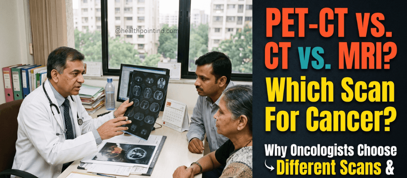
PET-CT vs CT vs MRI: What Each Scan Actually Finds
When cancer is suspected or confirmed, the scans start coming. A CT first, then perhaps an MRI, then somewhere along the way a PET-CT. To a family already absorbing difficult news, it can feel like repetition or indecision. It is neither. Each of these three scans answers a question the other two cannot, and understanding what your oncologist is actually asking at each stage makes the whole sequence considerably less bewildering.
The Simplest Way to Understand the Difference
Think of it as three separate questions.
A CT scan asks where is it and how big. It uses X-rays to build cross-sectional images, and it is fast, widely available and excellent at outlining structure. It will show a mass, measure it, and show its relationship to surrounding organs.
An MRI asks what exactly it is made of. It uses magnetic fields instead of radiation and produces far superior soft tissue detail. Where a CT shows a shadow, an MRI frequently shows what that shadow actually is, and how deeply it has invaded surrounding tissue.
A PET-CT asks: is it active, and is it anywhere else? This is the fundamental difference. PET does not primarily measure structure at all. It measures metabolic activity using an injected radioactive tracer, most commonly FDG, a glucose analogue. Cancer cells consume glucose faster than most normal tissue, so active disease lights up. The CT component performed in the same session supplies the map showing precisely where those active areas sit.
This is why a PET-CT can find disease in a lymph node that looks entirely normal in size on a CT, and why it can tell your oncologist whether a mass that is still visible after treatment is live tumour or just residual scar tissue.
When CT Is the Right Scan
CT is usually first, and for good reason. It is quick, broadly available, and surveys the chest, abdomen and pelvis in a single pass. For initial assessment when something abnormal has been found, for emergency situations, and for routine follow-up imaging during treatment, CT does the job well.
It is also the better scan for lung tissue specifically, where it outperforms both MRI and PET Scan for detecting small pulmonary nodules, and for assessing bone cortex detail.
Its limitation is that it judges lymph nodes largely by size. A node that is enlarged is treated as suspicious; a normal-sized node is treated as clear. Cancer does not always follow that rule, which is one of the main reasons PET-CT exists.
When MRI Is the Right Scan
MRI is the scan of choice when soft tissue detail determines the treatment decision.
Brain and spinal cord tumours are almost always assessed on MRI, since CT simply cannot resolve brain tissue adequately. Rectal cancer staging relies heavily on MRI to assess the circumferential resection margin, which directly decides whether a patient needs chemoradiotherapy before surgery. Prostate cancer detection has moved substantially to multiparametric MRI. Liver lesion characterisation, gynaecological cancer staging and musculoskeletal soft tissue tumours all sit in MRI territory.
MRI also involves no ionising radiation, which matters for younger patients and anyone needing repeated imaging over years.
The trade-offs are time, since scans run considerably longer, and the enclosed scanner, which some patients struggle with.
When PET-CT Is the Right Scan
Your oncologist orders PET-CT when the question is about disease activity or hidden spread rather than structure alone.
Initial staging in cancers that are reliably FDG-avid, including lymphoma, lung cancer, head and neck cancers, oesophageal cancer and melanoma. Here PET-CT frequently changes the stage assigned by CT alone, because it finds involvement that size-based assessment missed. That change in stage directly changes the treatment plan.
Treatment response assessment, particularly in lymphoma. A mass may remain visible on CT after chemotherapy while containing no live cancer at all. PET-CT distinguishes active disease from fibrotic residue, which no structural scan can do.
Recurrence detection when tumour markers rise but conventional imaging looks unremarkable.
Radiotherapy planning, where defining the biologically active tumour volume allows more precise targeting.
Its limitations are worth knowing honestly. Not every cancer is FDG-avid, and certain tumour types, including some well-differentiated thyroid cancers, low-grade prostate cancers and certain renal and neuroendocrine tumours, show poorly on standard FDG PET. For some of those, specialised tracers exist, including PSMA PET for prostate cancer and DOTA PET for neuroendocrine tumours.
PET also produces false positives. Infection, inflammation and recent surgery all raise glucose uptake, which is why a bright spot on PET is read in clinical context rather than treated as cancer by default.
A Real Sequence, in Order
Rajesh, a 54-year-old in Pune, was investigated for a persistent cough and weight loss. A chest CT showed a mass in the right upper lobe and two mediastinal lymph nodes of borderline size. Biopsy confirmed non-small cell lung cancer.
Before deciding between surgery and chemoradiotherapy, his oncologist ordered a PET-CT. It confirmed activity in the primary mass and in one mediastinal node, but critically showed nothing elsewhere in the body. An MRI brain was added separately, since lung cancer commonly spreads there and PET is less reliable for brain assessment because normal brain tissue consumes glucose avidly.
Three scans, three distinct questions, one treatment decision. None of them was redundant.
What Each Costs in Pune
Pricing reflects the technology involved rather than clinical importance.
CT scans through Health Point IND start from ₹1,499. MRI starts from ₹2,999. PET-CT starts from ₹11,999 to ₹13,999 against typical private rates around ₹23,000.
PET-CT costs substantially more for genuine reasons. The FDG tracer has a half-life of roughly 110 minutes, meaning it cannot be stored and must be produced fresh in a cyclotron on the day of your scan. The hybrid scanner is far more capital intensive than standalone equipment, and reporting requires nuclear medicine subspecialty expertise.
Most comprehensive health insurance policies cover PET-CT when prescribed by an oncologist for a diagnosed condition, though pre-authorisation is usually required before the scan rather than claimed afterwards.
What Preparation Each Involves
CT is the simplest. Fasting of around four hours if contrast is being used, and a recent kidney function test for contrast studies.
MRI requires no fasting for most protocols. All metal must be removed, and any implant, pacemaker or cochlear device must be declared when booking, since compatibility has to be confirmed in advance.
PET-CT scan is the most demanding. Six hours of fasting with plain water only, no tea, coffee, juice or chewing gum, because raised blood glucose competes with the tracer and degrades image quality. No strenuous exercise for 24 hours beforehand, since muscle activity takes up tracer. Stay warm, as cold activates brown fat, which also takes up FDG. The scan itself runs 20 to 40 minutes, but the full visit takes three to four hours, including the 60-minute rest period after injection while the tracer distributes.
Booking Cancer Imaging in Pune
Health Point IND books PET-CT, CT and MRI at accredited partner centres across Pune and PCMC. PET-CT needs booking, since tracer production must be scheduled against your specific slot.
Share your oncologist’s prescription on WhatsApp at +91 77964 00040, as it specifies the exact scan and tracer, and the team will confirm availability, protocol and a confirmed price before you travel.
Frequently Asked Questions
What is the main difference between PET-CT and a normal CT scan?
A CT scan shows structure, meaning the size, shape and position of a mass. A PET-CT additionally measures metabolic activity using an injected tracer, showing whether disease is biologically active and whether it has spread to sites that look normal on CT. PET-CT can identify cancer in lymph nodes of entirely normal size, which size-based CT assessment would classify as clear.
Why did my oncologist order a PET-CT when I already had a CT scan?
Because they answer different questions. The CT established where the disease is and how large. The PET-CT establishes whether it is metabolically active and whether it has spread elsewhere, which frequently changes the cancer stage and therefore the treatment plan. It is not repetition.
Can a PET-CT replace an MRI?
No. PET-CT and MRI assess different things, and MRI remains superior for brain and spinal cord tumours, rectal cancer staging, prostate assessment and soft tissue detail generally. Normal brain tissue consumes glucose avidly, which limits standard FDG PET for brain assessment, so MRI brain is frequently ordered alongside PET-CT rather than instead of it.
Does PET-CT detect all types of cancer?
No. PET-CT works best for FDG-avid cancers including lymphoma, lung, head and neck, oesophageal and melanoma. Some tumours, including certain thyroid, low-grade prostate, renal and neuroendocrine cancers, show poorly on standard FDG PET. Specialised tracers such as PSMA for prostate and DOTA for neuroendocrine tumours exist for several of these.
What does PET-CT cost in Pune compared to CT and MRI?
Through Health Point IND, CT runs ₹1,499 to ₹3,499, MRI ₹2,999 to ₹5,499, and PET-CT ₹11,999 to ₹13,999, against typical private rates of around ₹5,000, ₹9,000 and ₹23,000 respectively. PET costs more because the tracer must be freshly produced in a cyclotron on the day and the hybrid scanner is far more capital intensive.
Why is the PET-CT preparation so strict?
Because the FDG tracer is a glucose analogue, and raised blood sugar at the time of injection competes directly with it for uptake into cells, reducing contrast between cancer and normal tissue. Recent exercise and cold exposure similarly divert tracer into muscle and brown fat, creating areas that can obscure or mimic findings.
Does a bright spot on a PET scan always mean cancer?
No. Infection, inflammation and recent surgery all raise glucose uptake and can appear bright on PET. That is why a nuclear medicine specialist interprets findings in full clinical context, alongside your history and other imaging, rather than in isolation.
Three scans, three different questions, and a sequence that makes sense once you know what each one is for. Explore our PET scan, CT scan, and MRI scan services for details on each.
Important: This article is for informational and educational purposes only. It is not a substitute for professional medical advice, diagnosis, or treatment. Always consult your doctor or a qualified healthcare professional before undergoing any diagnostic test. For diagnostic test bookings in Pune, contact Health Point IND at +91 77964 00040 or visit https://www.healthpointind.com